
What is Odontoma?
Odontoma is a specific type of tumor that is benign and located in your mouth. It is made of all or some of the tissues that make your teeth. However, these tissues may or may not be arranged in the form of a tooth. It may not be visible to the naked eye. It is a mass of enamel, dentin, pulp and cementum that usually grows at the same rate as teeth. However, this mass is disorganized and any one of the components may become the dominant part.[1]
Odontoma is most often found in people around age 14 as a tooth that has not emerged. [3] More complex odontoma are found in males up to age 30. [7 ] Though more recent data indicates these tumors are found in both genders. [11]
Types of Odontoma
There are two general types of odontoma, some are more serious than others: Common odontoma and complex odontoma. These are further defined by specific components. They are described in part by their location and origin:
- Ameloblastic [2]
- Compound
- Radicular
- Temporal
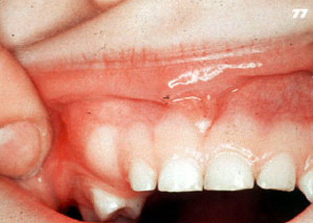
Picture : Odontoma
Symptoms of Odontoma
There are many symptoms of Odontoma but they are often overlooked or misdiagnosed. One that is serious is difficulty with swallowing. (dysphagia). If a tooth is delayed when it ought to be erupting, that is another sign that something is not right. If you have a lump in your gum that could be a symptom of odontoma. Some may be confused initially with an impacted wisdom tooth.
You could also have a primary tooth that is not coming out as it should. Also, if the bone beneath your tooth is enlarged, your dentist will notice this as a possible sign of odontoma.[4] These are some reasons why you need to keep a good connection with your dentist. That way the dentist becomes familiar with your teeth and oral conditions.
Diagnosis of Odontoma
Odontoma does not usually show external symptoms. These tumors are revealed when the x-rays are examined by the dentist. Although it is true that a delayed tooth or absent tooth may suggest there is a need for further examination. [5]
The presence of an a tumor of dental origin requires further examination to determine what type of tumor it is before further action is taken. In addition, a histological diagnosis of the tissues that were extracted provides valuable information to the dentist.
Odontoma Histology
No one really knows why an odontoma forms. The most likely reasons are trauma and/or infection at the site. Some dentists and researchers believe they are hereditary or they develop because of genetic mutations.[7] One example of an inherited syndrome is known as Gardener Syndrome. It is responsible for a wide range of tumors in the body, including occasional odontoma.[8] When examined at the cellular level, all of the dental tissues are found, but in an abnormal combination.
Pulp, dentin, enamel and cementum may sometimes resemble a tooth like structure in a compound odontoma. These denticles are found in a surrounding supporting layer of fibrous cells. Since it is decalcified, the enamel looks like spaces around the tiny tooth structures. [11] Looking closer, you can see the calicified material either as a solid mass or as multiple, small tooth-like bodies visible by x-rays. Because it is easily separated from its bony location it can be distinguished from other possible tumors.[7]
A complex odontoma has no specific sequence for all of the dental tissue. It does not resemble normal tooth structure. At the cellular level it appears as mostly tubular dentin that encloses hollow spaces. These circular spaces are decalcified but they once held enamel. On the edges there may be a thin layer of cementum which forms a capsule like tissue surrounding the mass. [11]
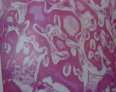
Histology Picture 1 : ODONTOMA (Complex) Histological features
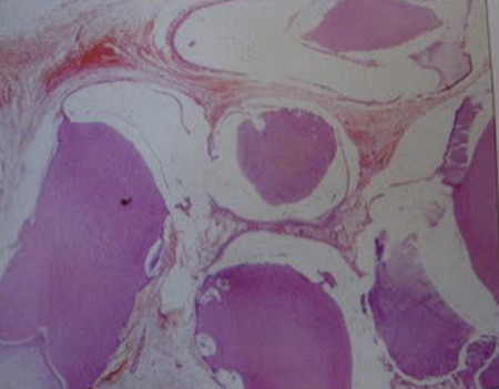
Histology Picture 2 : ODONTOMA (Compound) Histological features
CT Findings for Odontoma
CT findings refers to the images found by x-rays enhanced by computer tomography(a layered sectional view). These advanced technological images help the dentist see what is inside the tumor before engaging in surgery to remove it. They are each unique in formation and each arrangement of dental tissue can determine what kind of structure it has. Sometimes a complex odontoma can extend into the nasal cavity and require special attention.[9]
It is important to perform a complete examination beyond a simple x-ray to obtain this additional information. Sometimes magnetic imaging is recommended (MRI) to gain a thorough analysis of a complex mass.
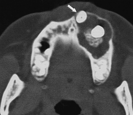
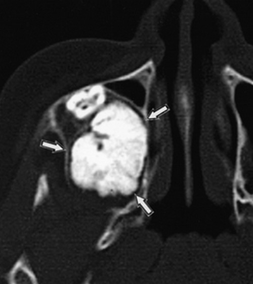
CT Picture 1 : Adenomatoid Odontogenic Tumor (Axial CT scan in left maxilla in 17 year old girl)
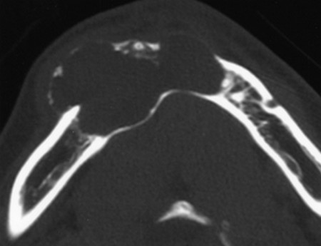
CT Picture 2 : Ameloblastic Fibroodontoma (Axial CT scan of right maxilla in 10 year old boy)
CT Picture 3 : Calcifying Odontogenic Cyst (Axial CT scan of mandible in 17 year old girl)
Treatment of Odontoma
The only real treatment of an these dental tumors is removal by surgery. An early discovery and treatment will be beneficial to the patient. It is a benign tumor made of dental tissue and it is a fairly simple extraction in most cases. A speedy recovery is generally expected. [6] Some complex tumors can result in complications after extraction. So it is essential to stay in contact with your surgeon. [7]
Research on Odontoma
How often are each of these dental abnormalities found? Which are more common? After reviewing 104 cases, 67 were identified as compound and 32 were diagnosed as complex. It is rare to find both types or multiple odontomas. Only 5 such cases were found in this study.[ 7]
Odontoma Video
References
- http://medical-dictionary.thefreedictionary.com/odontoma
- http://en.wikipedia.org/wiki/Odontoma
- http://www.rightdiagnosis.com/o/odontoma/intro.htm
- http://www.ncbi.nlm.nih.gov/pmc/articles/PMC2996496/
- http://www.ncbi.nlm.nih.gov/pubmed/11340730
- http://www.SciRP.org/journal/ojst/
- http://en.wikipedia.org/wiki/Gardner’s_syndrome
- http://www.thefreelibrary.com/Complex+odontoma+of+the+nasal+cavity%3a+a+case+report.-a0179614104
- http://www.ajronline.org/doi/full/10.2214/ajr.177.4.1770937
- http://dentallecnotes.
blogspot.in/2011/07/oral- pathology-lectue-note- odontogenic_23.html
Odontoma is caused by what?
How long it will take after Odontoma removal?