
What is Porencephaly Cyst?
Porencephaly is a very rare disorder of the central nervous system resulting in cerebrospinal fluid in cavities or cysts within the brain. The localized encephalomalacia (brain softening) is usually because of hemorrhage or inflammation of that portion of the brain. A less common form is inherited. [2]
It is possible that the fetus essentially had a stroke. It may develop before or after birth. It is usually found in infants or very young children. These cavities or cysts may be mild enough to not be noticed. Or they may be severe enough to show physical and mental disorders. [1]
Symptoms of Porencephalic Cyst
Some patients show no symptoms at all. Muscle spasms and seizures are common early signs. Delayed language development, muscle development, and mental incapacity are also common. Head size and shape may vary from very small (microcephalic) to expanding (hydrocephalic) due to the size and location and growth of cysts.[10]
Diagnosis of Porencephaly
Porencephaly is usually identified before the infant reaches the age of one year. Patients may live into their twenties in some cases.[1] The two types of porencephaly are developmental or congenital. Within the developmental type there are several additional sub categories. [10]
Porencephaly Radiology
To confirm the diagnosis of porencephaly cyst requires several tests and exams. The examination of the infant with magnetic resonance imaging, ultrasound and CT scans can reveal the presence of these porencephalic spaces or cysts, even in a pre-natal state after 20 weeks of gestation. [10]
Porencephaly MRI
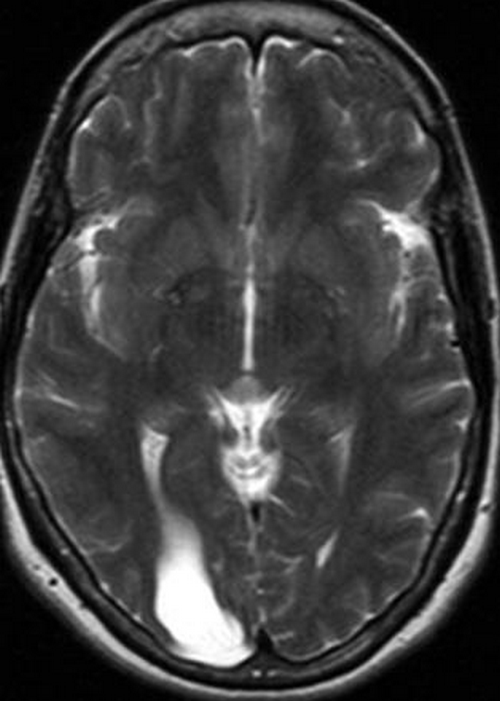
Picture 1 : MRI (T2)
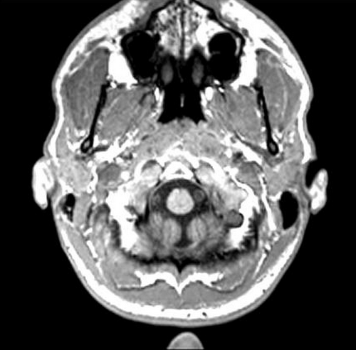
Picture 2: MRI (T1)
Porencephaly CT
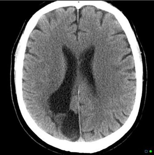
Porencephaly Cyst : Modality: CT (non-contrast)
Porencephalic Cyst
As early as 1859 porencephalic cysts were first seen and discussed by R. Heschl, though this precedes the development of the highly technical diagnostic tools available today.
In determining what kind of developmental cyst is present, physicians consider these differential imaging characteristics:
- Arachnoid cyst – extra axial, including gray matter beneath
- Interhemishperic cyst
- Neuroglial cyst – without communication between ventricles or subarachnoid space
- Holoprosencephaly – normal separation of the hemispheres has failed.
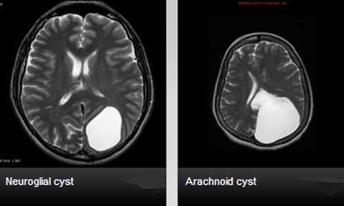
MRI : Difference between Neuroglial cyst and Arachnoid cyst
Modality: MRI (T2)
This is valuable information for the purposes of diagnosis. However, the prognosis and treatments remain essentially the same. The amount of impairment is dependent upon the size and location of the cysts. Treatment is essentially supportive for the infant. [10]
Porencephaly vs Schizencephaly
Schizencephaly and Porencephaly are both rare disorders in the brain. In fact, schizencephaly is a type of porencephaly. Patients have abnormal clefts or slits in their cerebral hemispheres. If the clefts are in both hemispheres, most likely they will have central nervous system dysfunction, minimal language skills and speech will be delayed.
If they have smaller splits or clefts in only one hemisphere they may have weakness on one side of the body and may not be mentally impaired. Other characteristics of schizencephaly include:
- mental retardation
- microcephaly,
- hemiparesis (weakness or paralysis which affects only one side)
- hypotonia (reduced muscle tone)
- quadriparesis (paralysis or weakness affects all arms and legs)
- hydrocephalus
- seizures.
This is a significant, though rare birth defect. One type of schizencephaly has a genetic origin. Other causes may be exposure to toxins, particular medications or cocaine use by the mother during pregnancy, or some unidentified external environmental abnormality or an unidentified vascular injury. Other signs indicate neurons have not succeeded in migrating to their proper place in the brain.
Treatments for patients with schizencephaly include physical therapy, medications to minimize seizures, and if necessary, a shunt to remove excess fluids from the brain. The prognosis for patients with schizencephaly will depend almost entirely upon the size and number of clefts and how much neurological damage has happened. [16]
The significant difference between schizencephaly and porencephaly that one is believed to be a primary developmental disorder where the neurons do not reach their destination, schizencephaly. The other, porencephaly, is a consequence of injury to brain tissue that causes it to soften or dissolve. These spaces can resemble each other. Some researchers theorize that they both have primary developmental disorders but at different stages of the development of the brain. It often requires magnetic resonance imaging (MRI) to examine the brain tissue along the edge of the cleft. In porencephaly it is usually white matter and scar tissue but in schizencephaly the cleft is lined with gray matter [17]
Porencephaly Cyst Treatment
Treatment for porencephaly is much the same as it is for schizencephaly. Since the degree of impairment depends entirely upon the size, location and number of cysts, and the patients are, for the most part, infants, physical therapy is provided. If epileptic symptoms are present, treatment for seizures is given. If needed, a shunt is provided to remove excess fluids from the brain. [5]
Research
Research is focused upon developmental issues and processes of the brain during the prenatal state. Much of the research investigates normal brain development, thus gaining insights into where things are different in these rare disorders. [2] In addition, since there are some cases of inherited porencephaly, there is genetic research underway to examine the genes involved.
It is described as an autosomal dominant trait, though these studies are in early stages. [6] A distinct genetic form of porencephaly occurs due to mutations of the COL4A1 gene. [9]