What is Gestational Trophoblastic Disease?
Gestational trophoblastic disease refers to different kinds of pregnancy-related tumors that develop the uterus. These tumors are usually rare and start as normal cells that develop in the placenta during pregnancy but would eventually grow out of control.
Trophoblasts refer to the outermost layer of cells that would form as the gestational trophoblastic tumors. It usually develops from the tissue that forms the wall of the placenta during the embryonic development.
Gestational trophoblastic diseases are usually benign tumors that can stimulate and emulate pregnancy complete with the development of an abnormal fetal tissue. Growth is usually localized in the uterus and does not invade body tissues or other body parts. However, there may be cases when malignancy may occur. Postmenopausal women may also be affected but this is usually a rarity.
Gestational trophoblastic disease may also be referred to as gestational trophoblastic neoplasia or tumors.
Different types of Gestational Trophoblastic Disease
There are five main types of gestational trophoblastic disease with one benign and four malignant tumors.
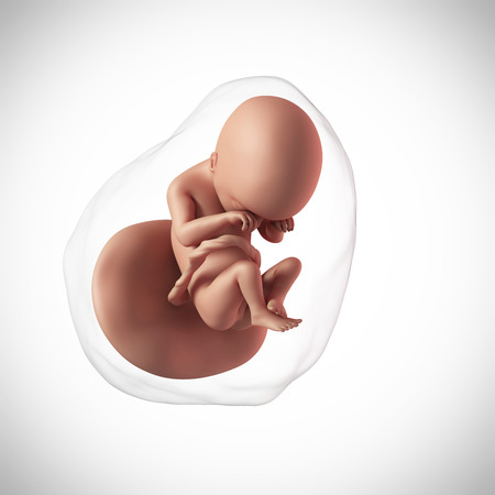
- Hydatidiform mole refers to the fertilization of an egg implanted into the uterus but with the failure of the chorionic villi to develop fully causing it to swell. The chorionic villi would form clusters similar to a bunch of grapes. The signs and symptoms of hydatidiform mole are similar to that of normal pregnancy but the pregnancy would not be viable. It may be classified as complete or partial hydatidiform mole and may also be referred to as molar pregnancy.
- Complete hydatidiform mole occurs when one or two sperms cells fertilize an egg cell without a DNA or nucleus. This type has a greater chance of developing into a choriocarcinoma.
- Partial hydatidiform mole occurs when 2 sperms fertilize a normal egg cell but develops with a trophoblastic tissue.

- Partial hydatidiform mole occurs when 2 sperms fertilize a normal egg cell but develops with a trophoblastic tissue.
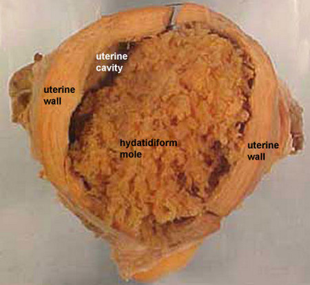
Picture 1 : Hydatidiform mole (Gestational Trophoblastic Disease)
- Invasive mole is also known as invasive hydatidiform mole or chorioadenoma destruens. It is a type of tumor growth that develops after conception into the muscular walls of the uterus. Invasive mole may spread and progress to other parts of the body like the vulva, vagina and even into the lungs. This usually occurs after the development of complete or partial moles.
- Choriocarcinoma refers to the tumor growth of fetal origins, usually that of the placenta. It is a germ cell tumor and has the tendency to invade and destroy the uterine wall as well as to metastasize through the lymph or blood vessels. Tumors of this kind are usually malignant and have the tendency to metastasize to other organs aside from the uterus.
Development of a choriocarcinoma usually occurs after a complete or partial hydatidiform mole, pregnancy, miscarriage or abortion.
- Placental-site trophoblastic tumor is a tumor growth that may secrete human chorionic somatomammotropin, a hormone that causes a positive test for pregnancy. This type of tumor develops when the placenta latches to the uterus lining. This usually occurs after a pregnancy, abortion or after the removal of a hydatidiform mole. Tumors of this kind do not usually metastasize but may invade the muscular walls of the uterus.
- Epitheloid trophoblastic tumor is almost similar to placental-site trophoblastic tumors. These tumors are exceedingly rare and are usually benign in behavior. The tumor usually develops after a full term pregnancy.
Risk factors associated with Gestational Trophoblastic Disease
There are various risk factors that could predispose a woman into developing gestational trophoblastic disease.
- Women under 20 years of age particularly those who are under 16 years old have six times chances of developing the condition.
- Women over 35 years of age especially those above 50 years old have one in every three chances of developing the condition.
- Previous gestational trophoblastic disease.
- Asian ethnicity.
- Women with Blood Type A or AB.
Signs and Symptoms
Although gestational trophoblastic diseases may exhibit signs and symptoms similar to pregnancy, there are various clinical manifestations that may indicate the development of tumor rather than that of pregnancy.
- Vaginal bleeding is the most common clinical manifestation that there might be a problem. It may start within the first trimester and may pass as brown blood clots.
- Anemia may result if there is excessive bleeding.
- Swelling and enlargement of the abdomen is also highly likely and may be faster than that of a normal pregnancy.
How is this disease diagnosed?
- Routine pregnancy tests are the usual diagnostic tools necessary in diagnosing gestational trophoblastic disease. These may include blood tests, ultrasound imaging and the usual diagnostic tests done after a miscarriage or abortion.
- Serum human chorionic gonadotropin hormone would be elevated leading physicians to first suspect that a woman may be pregnant.
- Biopsy may not be advised because of the risk of hemorrhage.
- Radiographic examination may be indicated to determine the location and size of the tumors.
Treatment Modalities
There are various treatment modalities for gestational trophoblastic diseases. The treatment undertaken would depend on the recommendation made by the physician as well as the decision of the patient. It is therefore important to have a broad understanding of all treatment options available.
- Surgery is the primary mode of treatment as this would allow the evacuation of pregnancy as well as the removal of the tumor. Suction curettage is the preferred method for evacuation while hysterectomy is the choice if future pregnancies are already ruled out by the patient.
- Chemotherapy is also indicated as a treatment of choice. Methotrexate and dactinomycin are the most common chemotherapeutic drugs used in the treatment. However, women who underwent chemotherapy are advised to avoid any pregnancy a year after the completion of treatment. These women are also predisposed to have an earlier menopause because of the medications used in chemotherapy.
- Radiotherapy may also be used if metastasis to other parts of the body has occurred.
There are cases when it is best to use two treatment modalities to be able to effectively eradicate the tumor and its growth.
Follow up check-up is necessary to ensure that there would be no further growth of any abnormal cells as well as to monitor the possibility of re-occurrence of the condition. Contraception may be indicated during the follow up period as pregnancy is discouraged during this time.
Gestational trophoblastic disease is a condition that may have symptoms similar to that of pregnancy. However, there would be instances that would greatly signify that the pregnancy is not a normal one. It is therefore best advised to have a physician examine the condition so that detection and treatment may be started promptly.