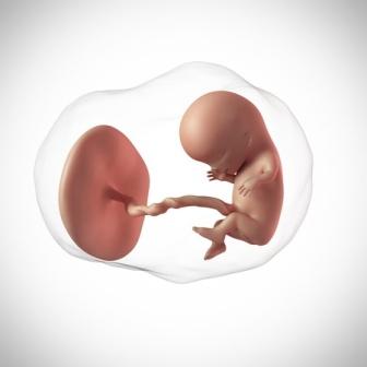
An ultrasound is a non-invasive diagnostic tool that is used to confirm pregnancy and to monitor the growth of the fetus throughout the nine months. The science behind pregnancy ultrasound is creating sound waves by a transducer which are reflected back from the uterus and baby. These waves are converted to visual images by a computer and are interpreted to get the exact details about your pregnancy and the baby.
How is Pregnancy Ultrasound Done?
An ultrasound is done by two methods, Transabdominal and Transvaginal.
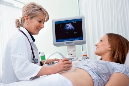
Transabdominal – ultrasound is more common. The technician will apply cold jelly on the abdomen and then with the help of a hand held probe (transducer), the jelly is spread across the tummy until the probe finds the exact location of the uterus for clear images of the baby. Images will be captured and seen on the screen as black and white moving objects. Most of the pregnancy ultrasound will be done by this method. In the early weeks of pregnancy around 11 weeks pregnant, the test will be done on a full bladder (you have to hold your urine till the bladder is full). The reason is, when the urinary bladder is fully dilated, the uterus which lies behind it is clearly visible.
Transvaginal – ultrasound is done around 6-8 weeks of pregnancy (after the onset of early signs of pregnancy). A cylindrical thin probe is covered with a condom and lubricated with jelly and then inserted into the vagina. The probe is moved in the vagina to check the uterus, tubes and surrounding structures. It gives accurate results in confirming early pregnancy (apart from the early pregnancy symptoms) or in case an ectopic pregnancy is suspected.
When a Pregnancy Ultrasound is Done?
A home pregnancy test is used to know if you are pregnant. But the confirmation of a viable fetus can be done only through an ultrasound. Pregnant women will undergo these scans at periodic intervals throughout nine months to monitor if the pregnancy is advancing normally. In a teenage pregnancy, an ultrasound greatly helps in detecting abnormalities that arise due to low age of the mother.
The first ultrasound is usually done when you are 10 weeks pregnant. In some ladies it may be done at 12 weeks pregnant. This first trimester scan is done to:
- Confirm an intra-uterine pregnancy
- To check for presence of heartbeat (absence of heartbeat is known as non viable pregnancy)
- To assess the number of fetus (single or multiple pregnancies)
- To calculate the due date.
- To rule out ectopic (tubal) pregnancy
A heartbeat can be detected as early as 6 weeks on a transvaginal ultrasound and by 12 weeks on a transabdominal.
The next scan done at 13 weeks pregnant is called the NT scan (nuchal translucency scan). Here the fluid collection at the back of the baby’s neck is measured. The results of this scan along with Triple marker test are used to assess the risk of chromosomal abnormalities like Down’s syndrome in the baby. During this scan, the sex of the fetus can be known in case the couple wish to find out.
The most important scan of pregnancy ultrasound is called as the Anomaly scan and is done when you are 20-22 weeks pregnant (second trimester scan). This is a lengthy scan and takes about 30 minutes. The technician will closely look at the basic anatomy of the baby. Measurements like BPD (bi parietal diameter), length of the humerus and femur are done and growth of the baby is assessed in number of weeks. The placenta, cervix and amniotic fluid are also studied in detail. Spina bifida and cleft palate can be picked up during this scan.
In the last trimester you may have a final scan with a color Doppler which is done mainly to check for the blood flow to the fetus and position of the placenta. It also shows the presentation of the fetus (breech or vertex) thus helping the doctor decide whether you can have a normal delivery or a C-section.
Risks and Benefits of an Ultrasound
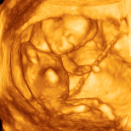
There are no known risks of an ultrasound as it is a non-invasive method. However, carrying out too many scans during pregnancy can lead to anxiety in the mother. These days, options for 3D and 4D ultrasound scans are also available which give a very clear picture of the baby. They are not mandatory or diagnostically better and results by the conventional ultrasound are considered equally accurate.